
|Articles|October 30, 2019
Should ob/gyns perform ultrasound examination at 35-37 weeks’ gestation?
Author(s)Ben Schwartz
Ultrasound examination at 35 to 37 weeks may reveal fetal anomalies that could not be observed in earlier ultrasounds, according to research recently published in Ultrasound in Obstetrics & Gynecology.
Advertisement
Ultrasound examination at 35 to 37 weeks may reveal fetal anomalies that could not be observed in earlier ultrasounds, according to research recently published in
Methods
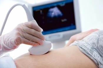
The prospective study included women attending for a routine hospital visit at 35+0 to 36+6 week’s gestation at one of two British hospitals between March 2014 and March 2019. The ultrasound examinations were performed transabdominally, using a 3-7.5 MHz curvilinear transducer, but in 2% to 3% of cases in which there were technical difficulties in obtaining adequate views, a transvaginal scan (3-9 MHz) was also carried out.
The research included 52,400 singleton pregnancies all of which had a previous scan at 18 to 24 weeks while 47,214 also had a scan at 11 to 13 weeks. The authors classified abnormalities according to the affected major organ system. Type and incidence of new abnormalities were also determined.
Results
At the time of third-trimester scan, median maternal age was 31.7 years (interquartile range, 27.5–35.4), median weight was 79.0 kg (interquartile range [IQR], 70.8 – 90.0), and median body mass index was 29.1 kg/m2 (IQR, 26.2 – 33.0). The majority of women in the study were white (74.9%) and the racial origin of the rest of the participants was black (15.4%), South Asian (3.8%), East Asian (2.0%) and mixed (2.9%).
The authors found that in the study population, incidence of fetal abnormality was 1.9% (995/52400), including 674 (67.7%) that had been diagnosed previously during the first and/or second trimester, 247 (24.8%) that were detected for the first time at 35 to 37 weeks and 74 (7.4%) that were detected for the first time postnatally.
The most common abnormalities observed for the first time in the ultrasounds at 35 to 37 weeks were:
- Hydronephrosis
- Mild ventriculomegaly
- Ventricular septal defect
- Duplex kidney
- Ovarian cyst
- Arachnoid cyst
Incidence of abnormalities seen at 35 to 37 weeks was 0.5% and those that were detected exclusively for the first time at the examination were ovarian cyst, microcephaly, achondroplasia, dacryocystocele, and hematocolpus. Incidence of abnormalities seen postnatally was 0.1% (74/52,400) and the most common were ambiguous genitalia or hypospadias, isolated cleft palate, and polydactyly or syndactyly. The authors noted that prenatal examination of the genitalia was not a compulsory part of the protocol.
Conclusion
The researchers said their findings indicate that a high proportion of fetal abnormalities are detected for the first time during ultrasound examination at 35 to 37 weeks’ gestation. They believe that incorporating ultrasound at that time in the gestational period could potentially improve postnatal outcome.
Newsletter
Get the latest clinical updates, case studies, and expert commentary in obstetric and gynecologic care. Sign up now to stay informed.
Advertisement
Related Content
Advertisement


Latest CME


Advertisement
Advertisement
Trending on Contemporary OB/GYN
1
Minipad-based HPV testing shows promise for cervical cancer screening
2
Kevin Ault, MD, on vaccines & autism, local government, HPV vaccine advocacy
3
Study data highlight trade-offs of testing for pregnancy too soon
4
Fionnuala McAuliffe, MD, highlights importance of timely breastfeeding discussions with patients
5