Press Release|Articles|September 16, 2025
‘Serotonin shield’: The placenta’s critical role in the health of babies

In a new study, Yale researchers show that the placenta regulates serotonin delivery to the fetus, contrary to past beliefs that it manufactures the hormone.
Advertisement
The placenta has long been thought to produce serotonin during pregnancy.
But in a new study, Yale researchers shatter the deep-rooted hypothesis — and show that the placenta doesn’t produce serotonin but instead regulates its delivery to the embryo and fetus. They found that serotonin comes from the pregnant parent, with the placenta acting as a “serotonin shield” that controls how much reaches the embryo and fetus.
The findings, published in the journal
“The placenta is in essence the ‘serotonin shield’ that regulates how much serotonin is ultimately delivered to the embryo and fetus, not the source of serotonin,” said
Often called a “happiness hormone,” serotonin regulates mood, so it’s often associated with the brain. In reality, less than 5% of serotonin is made in the brain, with 95% of it made in the gut. But serotonin does more than just regulate mood. It’s also a growth hormone. In the gut, it
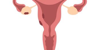
During pregnancy, serotonin also helps with growth: It travels into the placenta through a special protein known as the serotonin transporter (SERT) where it plays a critical role in the development of the embryo and fetus.
For the new study, researchers sought to better understand these relationships by using a pure source of placenta cells, unlike in previous studies that looked at either whole animals or isolated mouse placentas. To do so, they first
They also used another inhibitor called cystamine to block serotonylation, or the process by which serotonin is added to proteins like histone 3, which turns genes “on” and “off.” Again, that completely blocked the normal growth of the cells.
Blocking either SERT or serotonylation led to significant changes in gene expression of RNAs in the cytotrophoblasts, they found. Some genes — including ones involved in making, moving, and growing cells — became downregulated, or less active, when serotonin couldn’t enter the cell. Other genes — including ones that help cells stay alive and protect them — became upregulated, or more active. According to the researchers, these findings show that serotonin is critical for the growth of the cytotrophoblasts, the placenta, and by extension, the fetus.
Additionally, researchers discovered that the cytotrophoblasts don’t contain tryptophan hydroxylase (TPH-1), or the enzyme that makes serotonin, indicating the cells within the placenta can’t produce serotonin on their own.
“This suggests that factors that either inhibit serotonin transport through the placenta, or increase it, may have a significant impact on the placenta, embryo, fetus, and ultimately, the newborn and its brain,” Kliman said.
For example, Kliman says this explains why taking SSRIs — which decrease the levels of serotonin into the placenta — leads to smaller babies, and why, conversely, increased levels of serotonin may lead to bigger babies, with bigger brains, who may be at increased risk for developmental disabilities like autism.
Kliman and his lab have long investigated the link between placentas and children with autism, specifically the number of trophoblast inclusions (TIs) in the placenta. TIs are like wrinkles or folds in the placenta, caused by cells multiplying more than they should, typically only seen in pregnancies where there are genetic problems with the fetus.
This new study is the culmination of research
“This puts a big nail into the theory that vaccines cause autism,” suggested Kliman. “Autism, in essence, starts in the womb, not after delivery, and is most likely due to the genetics of the placenta and to a lesser extent, the maternal environment the placenta finds itself in.”
Kliman is also the director of the Reproductive and Placental Research Unit at YSM.
Other Yale authors include Gary Rudnick, a professor emeritus of pharmacology at YSM, and Seth Guller, a senior research scientist in obstetrics, gynecology, and reproductive sciences and director of the Gyn/Endocrine Laboratory at YSM.
This study was supported by grants from the Fulbright-Monahan Foundation, the University of Paris Cité, and the Reproductive and Placental Research Unit at Yale School of Medicine.
Advertisement
Related Content
Advertisement


Advertisement
Advertisement
Trending on Contemporary OB/GYN
1
GLP-1 RAs linked to broad menstrual adverse event signals in reproductive-aged patients
2
A discussion of STI rates, barriers, and home screenings with Damian Alagia III, MD
3
Q+A: Connor Frey, MD, on GLP-1 RAs and menstrual adverse event signals
4
CMS' obstetric G-codes cause confusion, perimenopause uncertainty, Zuranolone real-world data
5