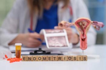
Noninvasive screening and diagnostic tool for endometriosis
Researchers from the Feinstein Institute in Manhasset, New York, found single-cell RNA sequencing to be an effective and noninvasive way to screen for and diagnose patients with endometriosis.
According to research in BMC Medicine, single-cell RNA sequencing could be a faster, more efficient way for ob-gyns to accurately screen and diagnose patients for endometriosis than surgery.
While nearly 1 in 10 women are diagnosed with endometriosis at some point during their reproductive years, the only way for ob-gyns to truly confirm the diagnosis is via excision laparoscopic surgery. This method is not only invasive, painful, and inconvenient for patients, but results can take days or weeks to process which could ultimately postpone treatment and delay symptom management.
Researchers from the Feinstein Institute in Manhasset, New York, used patient-collected menstrual effluent samples to conduct the first single-cell RNA sequencing comparison of endometrial tissues. They recruited 33 women aged 20 to 45 years old—with an average age of 33.6 years—via social media who were not pregnant or breastfeeding and were living in North America. Only women who were menstruating were included in the study.
Researchers divided the women into 3 groups—endometriosis, symptomatic, and controls. They categorized 11 women with histologically confirmed endometriosis—determined via excision laparoscopic surgery and pathology documentation—as
The endometriosis group included 11 women with histologically confirmed endometriosis—determined via excision laparoscopic surgery and pathology laboratory documentation.
The symptomatic group included 13 women who reported chronic symptoms consistent with endometriosis—recurrent dysmenorrhea, persistent abdominal bloating, dyspareunia, dysuria, and/or dyschezia—but did not have a diagnosis. The control group included 9 women who self-reported no gynecologic history that suggested a diagnosis of endometriosis, polycystic ovarian syndrome, or pelvic inflammatory disease.
Women in all 3 groups used an at-home menstrual effluent collection kit to get the menstrual effluent samples, which required the use of a menstrual cup for 4 to 8 hours on the day of their heaviest menstrual flow. One woman submitted her sample via saturated menstrual collection sponge, which had no significant impact on analysis or results.
Researchers used single-cell RNA sequencing to analyze endometrial tissue fragments in the samples and ultimately found a distinct phenotype of eutopic endometrial tissue shed into the menstrual effluent in patients with endometriosis when compared to control subjects.
They discovered a unique subcluster of proliferating uterine natural killer (uNK) cells in menstrual effluent tissues from the control group that were nearly absent from tissues of the endometriosis group and noticed a striking reduction of total uNK cells in the overall menstrual effluent of women in the endometriosis group.
After examining the endometrial stromal cells of women in the endometriosis group, researchers found a relative deficiency of progesterone-sensitive gene markers associated with endometrial stromal cell decidualization (e.g., IGFBP1, LEFTY2, LUM, CDN, etc.). They also found that the endometrial stromal cells of women with endometriosis were enriched in cells expressing pro-inflammatory and senescent phenotypes.
The study authors noted that an enrichment of B cells in the endometriosis group (p=5.8 x 10⁻⁶) raises the possibility that some women may have chronic endometritis—a disorder that predisposes patients to endometriosis.
The authors concluded that characterizing the endometrial tissues in menstrual effluent may be an effective screening method, especially in women with chronic symptoms suggestive of endometriosis. It may also help ob-gyns deliver accurate diagnoses quicker, which could begin to address the issue of delayed diagnosis endometriosis patients so often face. These findings may also lead to new diagnostic and therapeutic approaches for endometriosis and other related reproductive disorders.
Reference
1. Shih AJ, Adelson RP, Vashistha H, et al. Single-cell analysis of menstrual endometrial tissues defines phenotypes associated with endometriosis. BMC Medicine. 2022;20(1). doi:10.1186/s12916-022-02500-3
Newsletter
Get the latest clinical updates, case studies, and expert commentary in obstetric and gynecologic care. Sign up now to stay informed.


