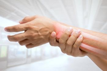
Osteoporosis in cirrhotics before and after liver transplantation
New research found that malnutrition in liver cirrhosis is significantly linked to osteopenia/osteoporosis for patients undergoing liver transplantation.
Bone mineral density (BMD) appears to be affected in patients undergoing liver transplantation, according to a study that found that malnutrition in liver cirrhosis is significantly linked to osteopenia/osteoporosis. The study in the
Methods
The study comprised 102 consecutive patients with cirrhosis referred for pre-liver transplantation (LT) evaluation at the
In 75 patients, serum-thyroid stimulating hormone (TSH), free triiodthyronine (T3), free thyroxine (T4), growth hormone (GH), cortisol, free testosterone, dehydroepiandrosterone sulfate, estradiol, interleukin-6 and tumor necrosis factor (TNF)-α were obtained.
Overall, 61 patients received a LT, 47 were followed for 1 year. During follow-up, nutritional status and BMD were assessed, and in 34,blood samples were analyzed.
Findings
Forty percent of LT candidates had osteopenia or osteoporosis and 38% were malnourished. Malnutrition was highly associated with osteopenia/osteoporosis: crude odds ratio (OR) = 3.5; 95% confidence interval (CI): 1.4 to 9.9.
In addition, the hip BMD Z-score decreased -0.25 (95% CI: -0.41 to -0.09) from baseline to 1-year post-LT.
High baseline TNF-α and high baseline cortisol levels correlated with a more marked decline in BMD (partial correlation (r) = -0.47; P < 0.05 and r = -0.49; P< .05, respectively).
The study “confirms that cirrhosis has a negative impact on BMD which seems to deteriorate during the waiting time up to and in the first year following LT,” the European authors wrote. “However, for patients with cholestatic etiology, there was no decrease in the hip BMD Z-score during follow-up, and patients with cirrhosis of other etiologies than cholestatic displayed a significantly greater decrease in hip BMD Z-score compared to those with cholestatic disease.”
Although malnutrition and low subcutaneous fat mass were associated with osteopenia/osteoporosis prior to LT, subcutaneous fat may actually protect against BMD loss, due to subcutaneous fat being linked to hormones such as estrogen and adiponectin. But the study found no connection between mid-arm muscle circumference (MAMC) and osteopenia/osteoporosis.
Conclusions
The authors noted that inflammation has recently been proposed as a potential major cause of malnutrition and of changes in body composition in patients with chronic inflammatory diseases. Chronic inflammation may also be associated with increased BMD loss, because TNF-α activates osteoclastic bone destruction and inhibits osteoblastogenesis in inflammatory diseases.
Furthermore, upregulation of interleukin-6 (IL-6) in cirrhosis can cause a prolonged acute phase response that subsequently could increase bone resorption.
However, the current study found a negative correlation between baseline TNF-α and BMD hip Z-score at 1-year post-LT, but no correlation with IL-6.
Similarly, an accumulated steroid dose of typically high doses of glucocorticoids after LT was not associated with post-LT BMD loss.
The authors advocate large cohort studies of patients with cirrhosis to confirm their findings and assess the potential role of malnutrition and systemic inflammation in the pathogenesis of osteoporosis in this population.
Newsletter
Get the latest clinical updates, case studies, and expert commentary in obstetric and gynecologic care. Sign up now to stay informed.


