
|Articles|September 1, 2007
Update on breech management
To minimize risks like birth-related trauma and asphyxia, current management stresses early recognition via ultrasound-which allows for an attempt to correct the breech-and reliance on C/S before onset of labor.
Advertisement
ACCREDITATION
This activity has been planned and implemented in accordance with the Essential Areas and Policies of the Accreditation Council for Continuing Medical Education (ACCME) through the joint sponsorship of CME2, Inc. ("cme2") and Contemporary OB/GYN. cme2 is accredited by the ACCME to provide continuing medical education for physicians.
cme2 designates this educational activity for a maximum of 1.0 AMA PRA Category 1 Credit(tm). Physicians should only claim credit commensurate with the extent of their participation in the activity.
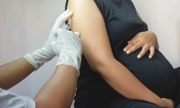
This cesarean breech delivery at term illustrates the importance of delivering the aftercoming head by force from behind. (Joel and Sharon Harris/Deborah Wolfe Ltd.)
TARGET AUDIENCE
Obstetrician/gynecologists and women's health practitioners.
EDUCATIONAL OBJECTIVES
After completing the following CME activity, the reader should be able to:
- Recognize the reasons for breech presentation and the benefits of early identification.
- Identify the confounding factors associated with adverse outcomes of breech presentation.
- Identify the preferred mode of delivery for the preterm breech weighing 1,000 to 2,000 g and the low birthweight breech (<1,500 g) and the risks associated with that choice.
- Summarize the appropriate circumstances for external cephalic version (ECV) as well as the approaches for ECV and its success rate.
TO EARN CREDIT FOR THIS ACTIVITY
Participants should study the article, log onto
DISCLOSURES
Editors Elizabeth A. Nissen and Paul L. Cerrato and Instructional Design Consultant Richard Currier, PhD, disclose that they do not have any financial relationships with any manufacturer in this area of medicine.
The manuscript reviewer discloses that he is a retained consultant for Adeza Biomedical.
Drs. Gimovsky and Bronshtein and Ms. Rosa disclose that they have no financial relationships with any manufacturer in this area of medicine.
RESOLUTION OF CONFLICT OF INTEREST
cme2 has implemented a process to resolve conflicts of interest for each continuing medical education activity, to help ensure content objectivity, independence, fair balance, and that the content is aligned with the interest of the public. Conflicts, if any, are resolved through a peer review process.
UNAPPROVED/OFF-LABEL USE DISCUSSION
Faculty may discuss information about pharmaceutical agents, devices, or diagnostic products that are outside of FDA-approved labeling. This information is intended solely for CME and is not intended to promote off-label use of these medications. If you have questions, contact the medical affairs department of the manufacturer for the most recent prescribing information. Faculty are required to disclose any off-label discussion.
Evaluating and managing the breech-presenting fetus involves wrestling with complex and ever-evolving issues-from medicolegal concerns to maintaining clinical skills. Our goal is to explore the differences between breech presentation and breech delivery and then look at the impact of the Term Breech Trial on your contemporary practice.
As you know, breech presentation is a longitudinal fetal lie in which the fetus' caudal end is the presenting part. Sometimes a fetus presents as a breech simply by chance. But otherwise, it's due to the relative amount of space a fetus has available to move within. Breech presentations may also be due to inherent maternal or fetal neurologic or orthopedic problems. Also influencing these issues are gestational age (GA), the presence of fetal anomalies, and the specific type of breech presentation (e.g., frank, complete with hyperextension, or footling).
Breech delivery may be vaginal or abdominal. Furthermore, vaginal delivery (VD) can be spontaneous, assisted, or by partial or total extraction. Whether the delivery of a breech fetus is vaginal or abdominal, the maneuvers must address a basic challenge: to accomplish the delivery of successively larger fetal diameters during birth (the opposite of delivery for a cephalic fetus). Inherent in breech delivery, these anatomical considerations raise concern for arrest or difficulty with the delivery of the fetal body or the umbilical cord, or both, before the head is completely delivered.
The greater risk of an adverse outcome seen with breech presentation compared to cephalic presentation is well-known and has been attributed to the relatively increased risks of prematurity, congenital anomalies, and birth-related trauma and asphyxia.1
Increased risk of prematurity
The incidence of breech presentation decreases with increasing GA. Spatial relationships, along with neuromuscular integrity and neurologic maturation, play key roles in fetal presentation. A longitudinal lie and cephalic presentation results from an adaptive process during which an active, normally proportioned, neurologically intact fetus (developing within an average volume of amniotic fluid) finds the best fit in the available intrauterine space.
Breech presentation is common in early pregnancy when the fetus is highly mobile within a proportionally larger volume of amniotic fluid. At 28 weeks, about 28% of fetuses are breech, falling to 3% to 4% at term.2 That translates into almost nine in 10 breech fetuses that convert spontaneously to cephalic prior to the onset of labor at term.
Changes in intrauterine volume that can potentially affect presentation in the preterm fetus can result from:
- uterine anomalies (bicornuate or septate uterus);
- space-occupying lesions (uterine leiomyomata);
- placental abnormalities (placenta previa, cornual placenta);
- parity (grand multiparity); and
- abnormalities in amniotic fluid volume (polyhydramnios).
Changes in fetal shape that can affect fetal presentation in the preterm fetus are:
- fetal anomaly (anencephaly, hydrocephaly, sacrococcygeal teratoma);
- extension of the fetal legs;
- fetal asphyxia;
- impaired fetal growth;
- neurologic impairment;
- short umbilical cord; and
- fetal death.
That's not to say the above conditions can't also affect term fetuses and contribute to a higher risk of breech delivery. There's a far greater risk, however, that the special conditions of prematurity will contribute to complications and death in the pre-term infant. About 2% of all live births in the United States occur at a GA of less than 32 weeks, and more than 12% occur before 37 completed weeks. Prematurity is the greatest single contributor to perinatal complications and fetal/neonatal death. Since the proportion of breech fetuses seen preterm is relatively larger than at term, it's not surprising that prematurity plays such a large part in adverse outcomes for breech-presenting fetuses.
Higher risk of birth trauma and asphyxia
In addition to newborn morbidity and mortality based on GA alone, preterm breech presentation significantly raises the risk of complications and death resulting from birth trauma and asphyxia. Vaginal breech delivery of the preterm fetus is linked with more cord accidents and a tendency for the relatively larger head to become entrapped in a partially dilated cervix. Further aggravating this tendency for entrapment is the higher number of breech presentations with incomplete flexion seen at earlier GAs.
THE PREFERRED MODE of delivery for the preterm breech is controversial, particularly for the very low birthweight (estimated fetal weight [EFW] <750 g) fetus.3-5 While cesarean section (C/S) is generally chosen for various reasons (such as the need for availability of neonatal intensive care), C/S early in the third trimester (or in the presence of premature rupture of the membranes [PROM]) can be technically more difficult than at term because of the thickness of the lower uterine segment.
IN THE PRETERM pregnancy (<36 weeks), the lower uterine segment is much narrower than at term, being on average only 4.0 cm (measured at the narrowest distance between uterine arteries) at 34 weeks.6 After 36 weeks, however, the lower uterine segment expands greatly. With a poorly developed, narrow lower uterine segment, a low transverse uterine incision may not be technically feasible or desirable and a transverse incision could increase the risk of birth trauma.6 Furthermore, a transverse incision can predispose to lateral extension of the incision, resulting in broad ligament or uterine artery laceration and subsequent maternal complications. A liberal use of vertical uterine incisions is indicated for the C/S delivery of the preterm or low birthweight (LBW) breech fetus, especially in the setting of PROM. In addition, the concurrent use of tocolytics has been recommended to facilitate extraction of the fetus at C/S under these circumstances, although this may increase the later risk of bleeding due to uterine atony.7
C/S OF THE PREMATURE breech fetus estimated to weigh between 1,000 and 2,000 g is widely practiced. In this weight range (roughly 26–36 weeks), the circumference of the fetal head exceeds the abdominal circumference, raising the risk of head entrapment during delivery and injury to the fetal cervical spine, abdominal viscera, or both.8 However, other risks complicate the choice between delivering by C/S versus VD: the significant risks of traumatic hemorrhage and perinatal depression for both the preterm breech fetus and the LBW (EFW <1,500 g) breech fetus.9 Respiratory distress, low Apgar scores, intracranial hemorrhage, birth trauma, and subsequent neurologic handicaps are more frequent among these small breech fetuses, regardless of the mode of delivery.
Increased risk of congenital anomalies
Serious congenital anomalies are also more prevalent in fetuses in breech presentation.10-12 It is therefore not surprising that perinatal complications and death due to congenital anomalies are greater for breech than cephalic fetuses. Congenital anomalies in both preterm and term breech presentation are up to three times more likely, when compared with fetuses in cephalic presentation at the same GA (Table 1).
THE MECHANISMS by which congenital anomalies are associated with breech presentation (and not breech delivery) are many and not entirely understood.13,14 Some posit that fetuses affected with CNS disorders or open neural tube defects are more likely to present as breech due to neurologic and neuromuscular impairment, which gets in the way of the fetus assuming a normal cephalic position.12,15
Congenital anomalies can also be associated with oligo/anhydramnios and interfere with rotation in the uterine cavity. The respiratory and GU anomalies that are associated with breech presentation would be expected to have abnormal amniotic fluid levels. Examples of these that might affect fetal presentation are lung hypoplasia, renal agenesis, Potter syndrome, and obstructive uropathy.
Fetuses affected with muscular disorders may be predisposed to improper fetal positioning due to hypo/hypertonia that impedes the fetus from moving within the uterus. Mothers with muscular disorders may also contribute a risk to fetal breech presentation: Researchers have found that maternal Duchenne muscular dystrophy (DMD) carriers have more fetuses in breech presentation regardless of whether the fetus is affected with DMD.15 The investigators hypothesize that DMD carriers have subtle changes in their uterine or pelvic girdle muscle tone that may increase the number of breech presentations.
Chromosomal anomalies/syndromes and genetic disorders associated with breech presentation are heterogeneous. Included in this category are chromosomal aneuploidy and other chromosomal anomalies (deletions, duplications, unbalanced translocations, etc.). The hypothesis is that chromosomal anomalies (including single-gene disorders and microdeletion syndromes) can lead to breech presentation due to the absence/excess of genetic material that encodes proper fetal positioning (neuromuscular development, renal development, etc.).
REGARDLESS OF THE various mechanisms thought to link congenital anomalies with breech presentation, it's important to focus on the specific malady and the prognosis of the affected fetus. When you find an abnormal fetal lie or presentation, a thorough search for fetal maldevelopment, including anomalies and IUGR, is in order.13 Thoroughly evaluate the affected organ systems (Table 1) by ultrasonography and magnetic resonance imaging (if indicated) and order prenatal genetic testing, where necessary. When a genetic cause is present, genetic counseling should permit further discussion that may influence the mode of delivery and neonatal management and outcome. In addition, counseling about these findings before delivery can prepare parents to be more realistic in their expectations.
External cephalic version (ECV)
Early identification of breech presentation allows you time to attempt to correct the breech. Both patient and physician benefit from recognizing breech presentation sooner, rather than later (during labor). Benefits are adequate time for counseling, sufficient time for informed decision-making regarding delivery, and the opportunity for a trial of external cephalic version.
ECV IS THE ONLY corrective treatment for a breech-presenting fetus. Conversion to cephalic presentation can be achieved in 60% to 70% of cases, lowering, but not eliminating, the breech presentation-related risks to the fetus and mother.14 Successful ECV occurs more often with multiparas, nonfrank breech presentations, amniotic fluid index (AFI) in the normal range for GA (10th to 90th percentiles), and in nonobese mothers. Tocolytics, amnioinfusion, and epidural anesthesia have been used to facilitate ECV.
AT PRESENT, we administer terbutaline subcutaneously (0.25 mg) after performing a non-stress test and prior to the attempt at ECV. When maternal diabetes complicates pregnancy, the tocolytic of choice becomes magnesium sulfate, given as a 4-g bolus. Although unusual, stillbirth (due to cord or placental accident) and other untoward events such as funic presentation and bony and peripheral nerve injury can result from ECV.16 That's why if labor is not immediately induced, we follow fetuses in whom ECV has been performed, whether successful or not, with twice weekly fetal testing. When breech presentation is diagnosed prior to labor and attempts at ECV fail, you can then consider a planned C/S or VD of a breech under nonemergent conditions and after a thorough discussion of the risks and benefits of either with the mother.
Breech delivery and the Term Breech Trial
Experience and data support the choice of C/S between 26 and 36 weeks in the interest of fetal/infant safety. As described previously, prior to 26 weeks, delivery by either route is fraught with risks, which makes a universal recommendation difficult.
EVEN MORE CONTROVERSIAL is breech labor and delivery after 36 weeks, when clinical considerations regarding a selective trial of labor versus universal C/S for breech delivery are subject to debate.17-21 The Term Breech Trial (TBT) reported a greater risk of immediate perinatal and neonatal death with planned vaginal delivery (PVD) than with planned C/S.19 However, follow-up reports at 2 years indicated that children born by planned C/S had no greater risk of death or neurodevelopmental delay than the PVD group.22 Likewise, there were no differences in maternal outcome between the groups at 2-year follow-up.23 In an evaluation of the TBT, Glezerman points out several concerns about adherence to protocol and the outcome measures the researchers relied upon.24
As a participant (MLG) in the TBT, it was clear that the investigators made a Herculean effort to study routine practices of breech delivery. The wide disparities in local practices clearly made adherence to the published protocol extraordinarily difficult to achieve and left meaningful results open to criticism. Local differences in selection criteria, augmentation of labor, management of the second stage, and immediacy of both emergent C/S and newborn resuscitation are also likely to affect both immediate and long-term fetal and maternal outcomes.
IN ADDITION, differences in sociocultural practices and liability concerns both affect the choices made and the evaluation of outcomes seen. Some believe that the TBT, despite admirable intentions, ultimately showed that breech management is an inherently complex issue, not amenable to study by simple randomization and that the TBT is not fully convincing in supporting the hypothesis under study. One researcher24,25 wonders whether the investigators' approach may have unintentionally skewed the outcome in favor of planned C/S (Table 2).
THE MAIN ISSUE is not whether you advocate a universal C/S, a selective trial of labor, or are uncertain.25,26 The TBT should be evaluated primarily by the methodology and outcome measures chosen. As with any rigorous study, these need to be appropriate to answer the clinical question. Thus, taken as a whole, it seems that the data reported by the TBT and others17-19,22-24 are consistent with either a trial of labor or universal C/S for term breech delivery.27
Our current management stresses the value of early recognition and reliance on C/S prior to the onset of labor. The liberal use of ultrasound in this setting has greatly increased our ability to diagnose breech presentation. Making the diagnosis of fetal malpresentation prenatally alleviates some of the stress from this diagnosis for both patient and ob/gyn. ECV can then be offered and-if acceptable to the couple-can be performed in a timely fashion. By a wide margin, most women will choose C/S. Vaginal delivery of the breech fetus currently is generally seen only in conjunction with emergent deliveries, twin gestation, intrauterine fetal demise, or with the extremely low birthweight (EFW <600 g) breech fetus.
UNIVERSAL C/S for breech delivery is rooted in the desire to avoid birth-related injury-and currently available data should be seen in that context. Adverse outcome for the breech presentation is often due to confounding factors that contribute both to breech and to newborn complications and death, such as prematurity, low birthweight, and congenital anomalies.
Reasons for the growing use of C/S stem from complex interactions in our cultural and social mores. The understandable but elusive desire to be 100% correct complicates the issue, especially regarding childbirth. Medicolegal concerns, cost containment, and attention to perinatal and maternal outcomes are relevant and evolving issues in contemporary practice, as is the maintenance of the necessary clinical skills.28
While not the final word on the subject, the TBT should promote further attempts to study and critically analyze direct clinical application of the evidence.25,26 A spirit of inquiry and the opportunity to improve the quality of health care need to be the "universal" approach.
REFERENCES
1. Cruickshank DP. Breech, other malpresentations and umbilical cord complications. In: Scott JR, Gibbs RS, Karlan BY, et al., eds. Danforth's Obstetrics and Gynecology. 9th ed. Philadelphia, Pa: Lippincott Williams & Wilkins; 2003:381-395.
2. Scheer K, Nubar J. Variation of fetal presentation with gestational age. Am J Obstet Gynecol. 1976;125:269-270.
3. Malloy MH, Oustad L, Wright E. The effect of cesarean delivery on birth outcome in very-low-birthweight infants. National Institute of Child Health and Human Development Neonatal Research Network. Obstet Gynecol. 1991;77:498-503.
4. Gravenhorst JB, Schreuder AM, Veen S, et al. Breech delivery in very preterm and very low birthweight infants in The Netherlands. Br J Obstet Gynaecol. 1993;100:411-415.
5. Wolf H, Schaap AH, Bruinse HW, et al. Vaginal delivery compared with cesarean section in early preterm breech delivery: a comparison of long term outcome. BJOG. 1999;106:486-491.
6. Morrison J. The development of the lower uterine segment. Aust N Z J Obstet Gynaecol. 1972;12:182-185.
7. Gimovsky ML, Vourlos D, Baldemero R. A way to minimize C/S trauma. Contemporary OB/GYN. 2005;50:32-33.
8. Tank ES, Davis R, Holt JF, et al. Mechanism of trauma during breech delivery. Obstet Gynecol. 1971;38:761-767.
9. Rayburn WF, Donn SM, Kolin MG, et al. Obstetric care and intraventricular hemorrhage in the low birth weight infant. Obstet Gynecol. 1983;62:408-413.
10. Brenner WE, Bruce RS, Hendricks CH. The characteristics and perils of breech presentation. AmJ Obstet Gynecol. 1974;118:700-712.
11. Braun FH, Jones KL, Smith DW. Breech presentation as an indicator of fetal abnormality. J Pediatr. 1975;86:419-421.
12. Reitberg C. Term breech delivery in The Netherlands 2006-Doctoral thesis, Utrecht University, The Netherlands.
13. Gimovsky ML, Petrie RH. The intrapartum management of breech presentation. Clin Perinatol. 1989;16:975-989.
14. Hofmeyr GJ, Kullier R. External cephalic version for breech presentation at term. Cochrane Database of Systematic Reviews. Available at http://gateway2.ovidweb.cgi Accessed May 15, 2002.
15. Geifman-Holtzman O, Bernstein IM, Capeless EL, et al. Increase in fetal breech presentation in female carriers of Duchenne muscular dystrophy. Am J Med Genet. 1997;19;73:276-278.
16. Bowes WA, Thorpe JM. Clinical aspects of normal and abnormal labor. In: Creasy RK, Resnik R, Iams J, eds. Maternal-Fetal Medicine: Principles and Practice. 5th ed. Philadelphia, Pa: Elsevier (USA); 2004:686.
17. Albrechtsen S, Rasmussen S, Reigstad H, et al. Evaluation of a protocol for selecting fetuses for vaginal delivery at term. Am J Obstet Gynecol. 1997;177:586-592.
18.Gimovsky ML, O'Grady JP, Morris B. An appraisal of CT pelvimetry within a breech management protocol. J Repro Med. 1994;39:489-491.
19. Hannah ME, Hannah WJ, Hewson SA, et al. Planned caesarean section versus planned vaginal birth for breech presentation at term: a randomized multicentre trial. Term Breech Trial Collaborative Group. Lancet. 2000;356:1375-1383.
20. Hauth JC, Cunningham FG. Vaginal breech delivery is still justified. Obstet Gynecol. 2002;99:1115-1116.
21. Munstedt K, von Georgi R, Reucher S, et al. Term breech and long term morbidity-cesarean section versus vaginal delivery. Eur J Obstet Gynecol Reprod Biol. 2001;96:163-167.
22. Whyte H, Hannah ME, Saigal S, et al. Outcomes of children at 2 years after planned cesarean birth versus planned vaginal birth for breech presentation at term: The International Randomized Term Breech trial. Am J Obstet Gynecol. 2004;191:864-871.
23. Hannah ME, Whyte H, Hannah WJ, et al. Maternal outcomes at 2 years after planned cesarean section versus planned vaginal birth for breech presentation at term: The International Randomized Term Breech Trial. Am J Obstet Gynecol. 2004;191:917-927`.
24. Glezerman M. Five years to the term breech trial: the rise and fall of a randomized controlled trial. Am J Obstet Gynecol. 2006;194:20-25.
25. Glezerman M. Reply. Am J Obstet Gynecol. 2006;195:1873-1874.
26. Ross S, Hannah M. Interpretation of the term breech trial findings. Am J Obstet Gynecol. 2006;195:1873.
27. Gimovsky ML. Breech Delivery. In: Queenan JT, Hobbins JC, Spong CY, eds. Protocols for High-Risk Pregnancies. 4th ed. Oxford, UK: Blackwell Publishing Inc.; 2005:555-558.
28. Deering S, Brown J, Hodor J, et al. Simulation training and resident performance of singleton vaginal breech delivery. Obstet Gynecol. 2006;107:86-89.
Advertisement
Related Content
Advertisement


Advertisement
Advertisement
Trending on Contemporary OB/GYN
1
When “free” isn’t free: the coverage gaps undermining breast cancer screening
2
ACOG releases own maternal immunization schedule, breaking from federal recommendations
3
Over half of elinzanetant's sleep benefit is VMS-independent
4
Stephanie Faubion, MD, MBA, on untangling sleep disorders from VMS in menopausal women
5