
|Articles|December 10, 2020
- Vol 65 No 12
- Volume 65
- Issue 12
Managing fetal sex discordance in cfDNA
This article presents a clinical algorithm and four cases to consider.
Advertisement
Introduction
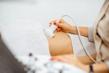
Cell-free DNA (cfDNA) screening for aneuploidy became available in the United States in 2011. Initially, it was offered as a screening option for women at increased risk of fetal aneuploidy, however, increasingly it has been offered as a screening option for all women.1,3,8 Performed as early as 9-10 weeks of pregnancy its high sensitivity (> 91%) and specificity (>99.6%) for trisomies 21, 18, and 13 from a single blood sample bolstered the test’s popularity over the traditional, sometimes multi-stepped serum analyte analyses.4 Additionally, unlike the alternative of serum analytes, cfDNA assesses the X and Y chromosomes with high sensitivities (> 97%) and specificities (>98%).2,4,5,7
As a consequence, fetal sex, if requested, is reported to the parents prior to the second trimester fetal anatomic evaluation.2,4,5,7 When cfDNA results for the sex chromosome complement are normal (XX or XY) but discordant with the appearance of the external genitalia on ultrasound, a range of etiologies can be responsible.
These may include disorders of external sexual differentiation due to either endocrine or sex chromosome aberration, disruption of normally developing external genitalia, or alternative sources of the cfDNA such as vanished twin, confined placental mosaicism and maternal medical conditions. Lastly, interpretation of both the common aneuploidies and the sex chromosomes lose specificity as fetal fraction declines with many laboratories not reporting results below fetal fractions of 4%.
We present a clinical algorithm to evaluate a pregnancy when discordance between the sonographic appearance of fetal external genitalia and cfDNA sex chromosome results is identified. The algorithm is supplemented by four cases, which exemplify these evaluations and the multidisciplinary approach needed.
Discordant cfDNA
Results for fetal sex and phenotypic appearance of fetal genitalia
Parents are often interested in finding out and confirming the sex of their unborn child. As cfDNA availability expands,6 more parents are given the information about fetal sex prior to the second-trimester anatomic survey.
Discordant results between fetal sex determined by cfDNA and the appearance of the genitalia on ultrasound inevitably will occur. Ongoing refinements in ultrasound resolution also aid not only the visualization of the fetal genitalia but also the more subtle findings of altered or under-development of the external genitalia.
As such, an algorithm to aid in the evaluation of these patients is useful. Our purpose is to provide an algorithm developed jointly among a multidisciplinary team for the stepwise assessment of such patients, augmented by clinical cases.
Four illustrative cases are presented that broadly represent the areas of potential concern to supplement the algorithms for differential diagnosis and management.
Cases
Case 1: Other source of DNA
This involves a 39-year-old Caucasian woman, gravida 2 para 1000, with no contributory past medical history with the exception that her first child died unexpectedly at 18 months of age with no definitive cause identified. This pregnancy was conceived by IVF with two embryos transferred.
At 6 weeks an ultrasound revealed a dichorionic-diamniotic pregnancy and ovarian hyperstimulation. Repeat ultrasound at 10 weeks revealed a singleton pregnancy with a second gestational sac containing a six-week sized embryo (CRL 2.5mm) without a heartbeat.
She underwent a 12-week ultrasound, which demonstrated a single normal-appearing fetus and a residual small second sac. Concurrent aneuploidy screening with cfDNA was performed and results reported disomy 13, 18, 21, and monosomy X and Y result. The fetal fraction was 11% and the sex was reported to the parents at their request.
Fetal anatomic survey at 18 weeks captured an image of normal female external genitalia; however, the discordance in sex assignment between the cfDNA was not recognized.
At birth, unexpectedly to the parents and the care team, the infant was noted to be phenotypically female. A karyotype was performed and resulted 46,XX.
The diagnosis is consistent with a presumed male, vanishing twin and underscores the significance for comparing the appearance of the external genitalia on ultrasound and the results based on cfDNA screening.
Case 2: Alteration of Normal Development
A 30-year-old Caucasian woman, gravida 3 para 0020, presented for care at 9 weeks gestation. She had no significant past medical history. She underwent aneuploidy screening with cfDNA at 11 weeks with results reported as disomy 13, 18, 21, and monosomy X, Y results. The fetal fraction was 4%.
Fetal anatomic survey at 19 weeks revealed phenotypically female genitalia. She underwent genetic consultation and declined diagnostic testing with amniocentesis. A repeat ultrasound at 28 weeks revealed severe hypospadias and short long bones with the femur lagging 3 weeks.
At that point, she agreed to further testing and an amniocentesis was performed with a normal chromosomal microarray consistent with 46, XY. A panel for 19 genes associated with disorders of sexual development disorders was sent and results were normal.
The baby was born at 37 weeks and was significantly growth restricted weighing 1928 grams (<1%). The newborn was noted to be phenotypically male, however, the phallus was short with normally descended testicles. A pelvic ultrasound was performed that did not reveal a uterus.
Normal testosterone and normal 7-dehydrocholesterol were noted. A clinical whole exome sequencing analysis was performed and no pathogenic variants were identified. In consultation with neonatology and pediatric urology, the diagnosis was most consistent with severe hypospadias and severe fetal growth restriction.9
Case 3: Disorders of Sexual Differentiation—Endocrine
A 39-year-old Caucasian woman, gravida 3 para 0010, presented for care at 8 weeks gestation. She had no significant past medical history. She underwent aneuploidy screening with cfDNA at 10 weeks gestation with disomy 13, 18, 21, and monosomy X, Y results. An anatomic evaluation at 13 weeks gestation was normal. A fetal anatomic survey at 19 weeks revealed a structurally normal fetus with normal-appearing female external genitalia.
Given the discordance between cfDNA results and phenotypic genital appearance, the patient was offered and underwent diagnostic testing that revealed a male karyotype 46, XY.
At birth, the infant was noted to be phenotypically female. Repeat karyotype on newborn peripheral blood was 46, XY. Pelvic ultrasound at birth revealed testicles within the inguinal canal and no uterus or ovaries.
In consultation with pediatric endocrinology and further testing, the findings are consistent with androgen insensitivity syndrome.
Case 4: Disorders of Sexual Differentiation—Sex Chromosome
A 29-year-old Asian woman, gravida 1 para 0 presented for care at 9 weeks gestation. She had no significant medical history. She had a normal 12-week ultrasound and aneuploidy screening with cfDNA, which was returned low risk for trisomy 13, 18, 21 and consistent with monosomy XY.
The fetal fraction was 11%. A sonographic fetal anatomic survey at 18 weeks revealed female external genitalia without identification of other structural abnormalities. She was seen by genetic counseling and a repeat cfDNA was performed with a similar low-risk, XY report.
Repeat ultrasound at 26 weeks and 5 days suggested a very small phallus with a bifid scrotum. She declined diagnostic testing. At birth, the infant was noted to be a slightly virilized female with a prominent clitoris.
An ultrasound at birth confirmed normal vagina, uterus, and ovaries. Neonatal peripheral blood karyotype was 46, XX and FISH with the Y chromosome specific SRY probe was notable for a positive signal in 4/100 cells.
The remainder of the 100 cells was negative for the FISH SRY probe. The infant had a buccal smear at birth to further evaluate mosaicism, which revealed 37.5% of cells with a single X (consistent with XY) and 62.5% with two X chromosomes (consistent with XX). Diagnosis is consistent with sex chromosome mosaicism.
Discussion
Ultrasound has a role in confirming concordance between the fetal sex reported on cfDNA and the external appearance of the fetal genitalia, if acceptable to the parents.
If there are inconsistent results further investigation is warranted and a systematic, multidisciplinary team approach involving communication and review by providers in imaging, molecular diagnostics, genetics, endocrinology, neonatology and urogenital subspecialists.
Genetic counselors and/or maternal-fetal medicine physicians often facilitate such team approaches. To guide this process, we propose a clinical evaluation for women when cfDNA sex chromosome results and the appearance of the fetal genitalia on ultrasound are discordant.
The conflicting results can be grouped into 4 broad categories 1) cfDNA not reflective of the fetus including vanishing twin, confined placental mosaicism, and maternal conditions such as transplant; 2) disorders of sexual differentiation due to a hormonal or sex chromosome aberrations; 3) alteration of normal external genitalia development associated with genetic syndromes or severe IUGR; and 4) analysis errors in the cfDNA interpretation associated with low fetal fractions.
A complete history and physical examination are always the initial step to guide further evaluation, including mode of conception and if by assisted reproduction, the number of embryos transferred.
A history of organ transplantation or transfusion, and exposure to medication, such as hormone therapy or virializing medications, should be evaluated. Family history should also be closely examined.
If any first trimester scans have been performed, these should be reviewed for the presence of multiple gestations or adnexal masses, for example, luteomas resulting in gestational hyperandrogenism.
The cfDNA result should be examined including the gestational age at which the test was performed (ideally greater than 10 weeks gestation) and the fetal fraction present.
Discussion with the reporting laboratory can help address both concerns related to a low fetal fraction or a disproportionate distribution of the sex chromosome signals suggestive of a mosaicism. Next, direct assessment of the fetal sex chromosome distribution by amniocentesis should be offered to the patient.
As with all fetal anomalies or concerns for underlying genetic syndromes, microarray can be performed on fetal DNA from the amniocentesis which can evaluate for microdeletion/duplication or low-level mosaicism.
Given the increase in accessibility of whole DNA sequencing and decrease of cost, whole exome sequencing can be pursued if additional fetal anomalies are present. Further investigation for Y chromosome-specific material with a SRY probe may be helpful to evaluate sex chromosome anomalies including translocation to X chromosome or autosome in XX individuals or mosaicism.
Further analysis for a single-gene condition causing a disorder of sexual differentiation encompasses both endocrine abnormalities such as congenital adrenal hyperplasia, and androgen insensitivity as well as a wide range of genetic syndromes, which impact external genital development.
Both the endocrine anomalies and the genetic syndromes can be present without other notable fetal anomalies detected by ultrasound.
As broad panels can include as many as 80 disorders, close communication with the DNA molecular lab can help refine the order of testing. For example, if the fetal karyotype is known, specific XX and XY panels may be available, including rapidity of results and cost.
A detailed sonogram should be performed by an experienced imager and include a comprehensive evaluation of the fetal anatomy because different constellations of findings may be associated with a variety of chromosomal and syndromic conditions.
Patients should understand that in some instances, detailed assessment of the external genitalia may not reveal changes until later in the third trimester.
The maternal adnexa should be evaluated for ovarian masses because on occasion certain types of lesions such as luteomas may be associated with virilization. Attention should be paid to fetal growth, as this may be a sentinel finding in identifying an underlying disorder.
Establishment of a specific prenatal diagnosis provides the mother with options for her pregnancy including obtaining the most accurate information from a multi-disciplinary group of subspecialty consultants, appreciating the underlying identified condition, the neonatal and pediatric care, and outcomes.
However, whether a definitive diagnosis is reached or not, the prenatal multidisciplinary team should include neonatology and as needed, individuals with expertise in the care of patients with urogenital differences. In all cases, there should be a thorough pediatric evaluation with physical exam at the time of birth.
Often this evaluation will include ultrasound imaging for presence or absence of pelvic organs that can further refine the diagnosis.
Further cytogenetic and molecular studies can be obtained if not performed previously. Based on these findings, the multidisciplinary team can direct its focus to endocrine evaluations, and approaches for urogenital care.
As more women use cfDNA for diagnosis, fetal sex will often be established prior to the second trimester anatomic survey. In these cases, the anatomic ultrasound can confirm concordance between the results of the cfDNA with the appearance of fetal external genitalia.
If discordance is present, further evaluation and a multidisciplinary team approach is warranted. The range of possible explanations is broad, with some explanations being clinically irrelevant to the fetal and newborn outcome while others represent significant medical conditions.
A thoughtful, stepwise approach to counseling and further evaluation should be undertaken to provide a diagnosis if possible, develop plans for evaluation of the neonate and the early incorporation of pediatric subspecialties as appropriate.
References
- Bianchi DW, Parker L, Wentworth J, et al. DNA Sequencing versus standard prenatal aneuploidy screening. N Engl J Med. 2014; 370:799-808.
- Bianchi DW, Platt LD, Goldberg JD, Abuhamad AZ, Sehnert AJ, Rava RP. Genome-wide fetal aneuploidy detection by maternal plasma DNA sequencing. MatErnal BLood IS Source to Accurately diagnose fetal aneuploidy (MELISSA) Study Group. Obstetrics and Gynecology. 2012; 119:890–901.
- Cell-free DNA screening for fetal aneuploidy. Committee Opinion No. 640. American College of Obstetricians and Gynecologists. Obstet Gynecol. 2015;126:e31-7.
- Gil MM, Quezada MS, Revello R, et al. Analysis of cell-free DNA in maternal blood in screening for fetal aneuploidies: updated meta-analysis. Ultrasound Obstet Gynecol. 2015; 45:249–66.
- Hill M, Finning K, Martin P, et al. Non-invasive prenatal determination of fetal sex: translating research into clinical practice. Clinical Genetics. 2011; 80 (1): 68-75.
- Gregg AR, Skotko BG, Benkendorf JL, Monaghan KG, Bajaj K, Best RG, et al. Noninvasive prenatal screening for fetal aneuploidy, 2016 update: a position statement of the American College of Medical Genetics and Genomics. Genet Med. 2016;18:1056–65.
- Norton ME, Jacobson B, Swamy GK, et al. Cell-free DNA analysis for noninvasive examination of trisomy. N Eng J Med. 2015; 372:1589-97.
- Screening for fetal chromosomal abnormalities. ACOG Practice Bulletin No. 226. American College of Obstetricians and Gynecologists. Obstet Gynecol. 2020; 136.
- Toufaily MH, Roberts DJ, Westgate MN, et al. Hypospadias, Intrauterine Growth Restriction and Abnormalities of the Placenta. Birth Defects Res. 2018; 110(2): 122-7.
Articles in this issue
over 5 years ago
Did L&D negligence cause urogynecologic injuries?over 5 years ago
State of the Industry 2020over 5 years ago
Lymphangioma circumscriptumover 5 years ago
The year in reviewover 5 years ago
Contemporary OB/GYN®: A look back to 2020over 5 years ago
A special thanksAdvertisement
Related Content
Advertisement


Advertisement
Advertisement
Trending on Contemporary OB/GYN
1
Subfecundity, not IVF, linked to elevated neurodevelopmental risk in offspring
2
Kate McLean, MD, MPH, explains microbial subtypes within bacterial vaginosis
3
Daré Bioscience launches prescription-free vaginal probiotic capsule for microbiome support
4
At-home pelvic ultrasound feasible, preferred over in-clinic care
5