
|Articles|June 14, 2011
Frequency of Uterine Malformations in a Restricted Gene Pool Community
Author(s)OBGYN.net Staff
This random retrospective cross-sectional study was preformed to determine the frequency of uterine malformation in restricted gene pool communities. In 4 groups of women desiring to conceive during their reproductive years, all women lives in lacrete, a community in north Alberta, Canada
Advertisement
Original Research by: Dr-Saad Ramzi Ismail
PHD-Public Health
MSc-Medical Ultrasound
BSc-Diagnostic Imagine
ARDMS, CARDUP, CRVS, CRGS, CAMRT
Ultrasound Supervisor
NWHC, High Level Hospital, Alberta, Canada
Box 1462, High Level,
Alberta T0H, 1Z0 Canada
August 2007
The author acknowledges no commercial affiliation or financial conflict of interest.
The author wishes to express his appreciation to Dr K. Hofmann, Dr P Hughes, Dr S Desilva, Dr Ali Cadili, Dr Bouta, Mrs. Valetta Lawrence, and Siti Arabiah. This paper is dedicated to my parents and my two brothers, who died in Iraq.
Abstract
This random retrospective cross-sectional study was preformed to determine the frequency of uterine malformation in restricted gene pool communities. In 4 groups of women desiring to conceive during their reproductive years, all women lives in lacrete, a community in north Alberta, Canada
Methods
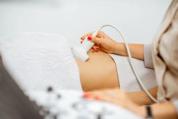
During2003-2006, eight hundred women from the community of LaCrete in northern Alberta (known restricted gene pool for over 500 years) were obtained. All subjects were scanned utilizing 2-D abdominal and transvaginal sonography. All gave informed written consent for participation in this study. The ethical committee of the education counseling service in High Level approved this study.
Results
156 patients (19.5% of the study population) showed some form of structural uterine malformation. This is 5.5 times higher than what is reported to be as the incidence in general populations in the literature. All 156 subjects were further investigated with MRI, 3-D sonography, hysterosalpingiography, Hysteroscopy, and laparoscopy. This testing revealed that 30 of these women had had no previous pregnancies or live births; 40 had one or more previous pregnancies and live births and more than 2 miscarriages; and 55 had with recurrent miscarriages. 95 women with recurrent and some miscarriages had more uterine malformation than the other groups, the result was tabulated according to the type of malformation and percentages in each group. The incidence in Group 3&4(infertile) was 60.8% and 39.2% in fertile group 1&2.The p <0.01 was significant at 95% confidence interval.
Conclusion
This study has provided new insights into the increasing number of uterine malformations in the population of lacrete. It was 19.5%, 5.5 times higher than that of general populations of (0.1 to 3.5%). Our study found a significantly higher prevalence of uterine malformations in a restricted gene pool community compared to the reported prevalence in the general population. This might indicate a direct link between lack of genetic diversity and the development of uterine malformations in women. A combination of TVS, hysterosalpingography, 3-D sonography, MRI, are necessary for the precise classification of uterine malformations.
Advances in Knowledge and Application to Patient Care
This study contributes to advances in knowledge as it shows that communities that practice endogamous marriages and lack of genetic diversity can see an increase the risk of uterine malformation to more than 5 times that of the general population. We can then educate these communities to practice a wider choice of marriage selection between different families. Moreover, not restrict them to few in order to lower the genetic risk linked to recurrent miscarriages.
The application to patient care is in the knowledge that gain by the researchers and doctors that can be applied both to the correct diagnosis of uterine malformation and to the management of patients with these abnormalities. Doctors and sonographers can use this study to improve skills in the difficult diagnosis of unicornuate uteri. The ability to diagnose uterine malformation has been improved by new imaging techniques.
Introduction
Little, if any, information exists in the literature regarding incidences of uterine malformation in restricted gene pool communities. Such information would be invaluable to the gynecologists and other healthcare professionals involved in the care of these patients. La Crete is a small town located 56 km southeast of High Level in Northern Alberta, Canada. The estimated population of LaCrete is 3000, almost 100% of Mennonite origin. The entire population is descendent from only 26 different families. These 26 families have been sharing genetic material through marriages since the 1500s (over 500 years) 1` In fact, marriages outside these 26 known families are estimated to occur in LaCrete in only less than 1% of cases. They DO not marry a first or second cousin 1. The term "Uterine malformation" is a general term encompassing a group of congenital anomalies affecting the female genital system. Such anomalies result from four main defects in the development and/or fusion of mullerian or paramsenephric ducts during fetal life 2, 3. The importance of diagnosing uterine malformations lies in their association with miscarriage and infertility 4. Some mullerian defects can have normal reproductive outcome5. Only those women with two or more pregnancy losses were defined as infertile6. The true incidence of mullerian defects in the general population is not known since they tend to be a symptomatic, usually only discovered accidentally, or in relation to pregnancy complications. Generally, 0.1 to 3.5% is the number accepted by many authors and these numbers changes according to each type of malformation 4, 7. Almost all structural uterine malformations are due to errors of development occurring in very early pregnancy. Embryo-logically in female fetus caudal ends of the two mullerian (paramesonephric) ducts fuse to form the uterus, cervix and upper vagina, whereas the un- fused cranial ends form the paired fallopian tubes 3, 8. Different types of fusion may occur including malformation of either or both the uterine body and uterine cavity, with subsequent impaired reproductive performance 9 10, 11. Such malformations affect negatively the reproductive performance of the uterus leading to increasing incidence of abortion and preterm delivery in women with uterine anomalies such as unicornate, bicornate, didelphys and septate uteruses 12,13, 14, 15 & 16. This is seen clinically as an increased rate of primary infertility (failure to conceive) or more often as an increase rate of early pregnancy loss (impaired implantation and early development). Later in pregnancy, unsuspected uterine malformations may present as impaired intrauterine fetal growth due to abnormal placentation, or abnormal fetal positioning related to mechanical factors in the shape of the uterine cavity. Labour, delivery, and third stage problems may occur related to in-coordinate uterine muscular activity12-16. A didelphus uterus is very rare anomaly and it can lead to a pregnancy failure17. Mullerian defects are associated with increased incidence of congenital urinary malformation. These incidences noted more in the unicornuate and bicornuate uteruses .18,19 Raga (1995) reported that the overall frequency of uterine malformation was 4.o%; and there was higher incidence of miscarriages amongst patients with septated forms of uterine malformations, it was recorded at 33.6% 20. Recurrent abortion, i e consecutive abortion on three occasions is less common and the etiology is unknown 21-25. Some of the associated factors include Genetic factors such as, thrombotic disorders, maternal medical disorders, and congenital malformation of the uterus. The relative contributions of these are unknown.20-25. Most studies demonstrate a spontaneous miscarriage rate of 10-15% however; the true rate of early pregnancy loss is closed to 50% because of the high number of chemical pregnancies that are not recognized in the 2-4 weeks after conception 26. Most of these pregnancy failures are due to gamete failure 26,27. The term “infertility” has a specific definition, which is the inability to achieve pregnancy despite unprotected regular and adequate intercourse over 12 months period. It has been wrongly used in many literatures to describe women with recurrent abortions 27. 3-D sonography is very sensitive and it should be utilized for the diagnosis of uterine malformation28. Unicornuate uterus is associated with poor reproductive outcome and high abortion rates up to 22% reported in the first trimester29. It is important to be precise in the diagnosis of uterine malformation as it can change the management of the treatment 30. (See
The major disturbances in the development of the Mullerian ducts (2,4&8) (See Diagram #1)
I. Failure of one or more Mullerian ducts to develop due to agenesis; unicornuate uterus without rudimentary horn.
II. Failure of the ducts to canalize (unicornuate uterus with rudimentary horn without proper cavities).
III. Failure or abnormal fusion of the ducts (uterus didelphys, bicornuate uterus).
IV. Failure of re absorption of the midline uterine septum (septate uterus, arcuate uterus)
Methods
This was a random cross-sectional and retrospective study of 800 women in Lacrete, carried out during 2003-2006. The women mean age was 31 years. All were referred for sonographic examination for a variety of reasons such as previous miscarriages, early pregnancy, infertility, pelvic informatory disease, or to rule out polycystic disease. All were scanned utilizing2-D abdominal and transvaginal sonography (TVS). The objective was to determine the frequency of uterine malformation among women in restricted gene pool communities. All subjects gave informed written consent to view their charts and conduct the research and to review the reports from other modalities. The ethical committee of the education counseling service in high-level hospital approved the study protocol in December 2003. A sub-group of 156 patients was identified with suspected uterine malformations; of these 78 (50%) belonged to other 22 families in the community. analysis of the patients was done according to the recurrent pregnancy loss status. All 156 women were referred for further diagnostic investigation to reach definite diagnosis Including Hysterosalpingiography, MRI, 3-D sonography, Hysteroscopy, and laparoscopy. Each modality has its own criterion of diagnosis according with the Canadian association of medical radiation therapy and the Canadian society of diagnostic medical sonographers (
1. Hypo-plastic uterus (vaginal, cervical, fundal, tubal and combined).
2. Unicornuate uterus (communicating rudimentary horn, without communicating rudimentary horn, with a rudimentary horn and without cavity, without horn.
3. Didelphus uterus
4. Bicornuate uterus (complete and partial)
5. Septate-uterus (complete and partial)
6. Arcuate uterus.
7. DES Drug Related malformation. Therefore, it is not genetic malformation.
Statistical Analysis
This was performed using chi-square X2 mean tests (which is the degree of freedom) utilizing SPSS Test package, Chicago, IL, USA); P< 0.01 was statistically significant at 95% confidence interval.
Results
The result of 156 women was tabulated according to the uterine malformation (
Discussion
The true incidence of uterine malformation in the general population is hard to determine as most data were derived from studies of patients with reproductive problems, and accurate diagnosis and complete assessment of the uterine malformation has not always been performed. The frequency of uterine malformation in most literature is 0.1% to 3.5 4,7.There is no studies in the literatures in regards to the frequency of uterine malformation among women belonging to restricted gene pool communities. Part of the problem lies in defining “restricted gene pools” and in quantifying this phenomenon. The entire populace of the study population (Lacrete) consisted of only 26 distinct families with a long tradition (over 500 years) of marrying only within these families. Of the 156 subjects included in this study, 78 (50%) belonged to only 4 of those families. Higher frequencies of uterine malformation are associated with higher rate of reproductive failure. No rates of uterine malformation for different ethnicity are available in the literatures. The worse outcomes recorded in this study for miscarriages were patients with septated uteruses (complete& partial forms) 41 patients, 26.8%. 26 of septated uteruses (both forms) were noted in-group 3 and 4,this is agreeable with the literatures dealing with septated forms of uterine malformations in relation with higher incidence of miscarriages. Group 3 had 7.6% Unicornuate uteruses; group 4 had 10.2% unicornuate uteruses and they have second higher incidence of miscarriages. Followed by patient’s in-group 3 had 5.1% and group 4 had 8.9% of bicornuate uteruses? This might indicate that this kind of malformation are most likely affect the reproductive outcome and increases the chance of miscarriage 12,13 & 20. There is an increase incidence of unicornuate and bicornuate uterus in the infertile population in-group 3 and 4 in comparison with fertile patients in-group 1 and 2. This support the study done by (Green and harris, 1976 and Acien 1993) 13,14 on a poor reproductive outcome associated with a bicornate uterus.156 patient with suspected uterine malformation diagnosed utilizing transabdominal and TVS were sent to have Hyesterosalpingiography to confirm the diagnosis. 22 of them had an MRI to further prove the diagnosis in the subseptate and arcuate uteruses which is difficult to diagnose using other modalities. 40 patients were sent to have 3-D sonography. 16 patients were sent to have hysteroscopy and 5 sent to have laparoscopy to treat endometriosis. Four cases of uterine malformations were missed using 3-D sonography due to the presence of fibroids causing the uterine wall to appear convex. The 3-D sonography imaging showed 90% sensitivity and specificity and was less sensitive in the presence of uterine lesions such as fibroids and polyps. MRI in another hand showed 100% sensitivity and specifity it showed more accuracy, precision and reliability, it should be the modality of choice after the initial diagnosis with TVS. Six Patients had problem with this imaging modality due to the claustrophobic effect. MRI is expensive procedure and the waiting list can reach to 6 months in Canada. TVS with color flow can differentiate vascular from non-vascular uterine septation. Unicornuate uteri have the highest rate of cervical shortening and this could affect the reproductive performance. In addition, it is helpful tool for the sonographer to correlate the shortening of the cervix to the unicornuate uterus, as group 3 & 4 had 85.7% cervical shortening. The least frequent malformation noted was the didelphus uterus 2 in 800 patients, this finding indicate that this type of rare malformation is very high in this community knowing that the incidence of didelphus uterus is rare 1/3000 17. The incidence of uterine malformation for fertile patients recorded at 39.2% and in the infertile patients at 60.8%. This indicates that it is common to have uterine malformation but it increase in the infertile women by 21.6% in this study. TVS, 3-D sonography and Hysteroscopy were all sensitive but MRI and hysteroscopy were more specific in detecting focal endometrial polyps. Laparoscopy is invasive procedure and used for the treatment of 5 patients with endometriosis that was discovered by MRI. 42 retroverted uteri were diagnosed using TVS in the infertile group 3&4.This might indicate a relationship between retroverted uteri and infertility but this is not relevant in this study, as the only link is where women have a fixed retroverted uterus on pelvic examination. This might indicate either past serious infection with scarring causing fallopian tube blockage, or due to endometriosis scarring. The methods of choice to detect uterine malformation should be discussed between the physicians and the radiologists to reach the best available method with less invasiveness to the patient. This study showed that the use of 3-D, TVS and transabdominal sonography should be utilized first followed by more expensive modality such as MRI. HSG is invasive method and can provide a faster diagnosis with out the claustrophobic effect of the MRI to some patients.
Two women with unicornuate uteri had children; this might indicate that women with this type of malformation can be pregnant and have successful delivery. 11 hypo-plastic uteri were recorded in this study 5 in the fertile group and 6 in the infertile group more studies need it to be done to know the effect of this malformation on fertility and increase miscarriages. Double the numbers of bicornuate uteri were confirmed in the infertile than the fertile group. This might indicate a relationship between these types of malformation, infertility and increase in miscarriages.
Sixteen septate and sub-septate uteri were found in-group 1&2 compare to 26 were found in-group 3&4.Again this might indicate the relationship between these types of malformations, infertility and increase in miscarriages, this support many literatures regarding the relation of the septated forms of uterine malformation to early miscarriages. The size of the septation may influence the reproductive outcome. There were 22 arcuate uteri in the fertile group compare to 11 in the infertile group, this might indicate that this type of malformation it does not effect the uterine productive performance, this type of malformation is difficult to diagnose utilizing TVS or trans abdominal sonography and it is better diagnosed using MRI, 3-D sonography, or laparoscopy. DES (diethylstilbestrol) a synthetic form of estrogen that was prescribed between 1938-1971 to help women with certain complications of pregnancy .It was linked to clear cell denocarcinoma of vagina and cervix of the unborn female fetous in women who are taking this drug. Therefore, DES is not related to uterine malformation classifications but sonographers should know the history of such drug effects. The urinary anomalies are not discussed in detail in this paper despite its relevancy as it is not the topic of this research and it will be discussed farther in separate study project.
Limitation and Farther Studies
The limitation in this study was that we did not use 3-D sonography for all patients; we should utilize it, as it is accurate than other modalities but less sensitive in the presence of fibroids. Moreover, we did not send all the 800 women to perform the special procedures such as MRI, HSG, Hysterscopy, and Laparoscopy. We could miss many uterine malformations by using transabdominal / TVS alone.
Future studies should focus on the distributions and the percentages of each anomaly and correlate them with the general populations. Correlation with different communities with restricted gene pool or of that practicing co-sanguinity might be beneficial.
Conclusion
Analysis of 800 fertile and infertile patients in the Lacrete female population has provided new insights into the incidence of uterine malformation in this population, which practice endogamous marriage. Lacrete has an incidence of 19.5%, 5.5 times higher than the general populations. Arcuate, and retroverted uteri may be irrelevant to the reproductive performance of women. Septated versions of the uterine malformation and the bicornuate uteri are increases in the infertile groups, depending on the size of each uterine horn. Different diagnostic methods should be used to classify each type of uterine malformation for precise classification of these malformations. A combination of 3-D, TVS sonography (non invasive technology) and HSG (invasive) seem to be necessary and less expensive than MRI technology. Uterine malformations are relatively frequent in fertile women; but more frequent in infertile groups, and in communities with restricted gene pool. These malformations can impair the pregnancy outcome. The uterine malformations are more common in women with recurrent miscarriages. The reproductive performances of, unicornate, septated and didelphus uteri were poor. Mullerian defect can permit normal delivery depending on the type of the uterine malformation.
1-hypoplasis /agenesis uterus, it can be vaginal, cervical, fundal, tubal or combined agenesis.
II-Unicornate uterus, it can be communicating, non-communicating, with no cavity and with no horn.
III-Didelphus uterus, it is with 2 vagina, 2 cervixes, 2 cavities and 2 horns.
IV-Bicornate uterus, it can be complete or partial.
V-Septate uterus, it can be with complete septation or partial septation (sub septus).
VI-Arcuate uterus, it is like normal uterus with fundal or myometrium indentation.
VII-this is caused by drug changes and not genetics, there for it is not classified as uterine malformation.
Courtesy of RSNA-
Letter of permission was granted by the radiological society of North America to use these figures.
Hystrosalpingography (HSG) the arrow shows the right uterine horn and missing left horn of the uterus.
This type of malformation called the Unicornuate uterus
Figure 2
HSG. Bicornuate uterus, the arrow shows the division of the uterine horns
Figure 3
HSG. The arrows shows Didelphus uterus. Each horn with complete vaginal vault and cervix to each side of the uterus
Figure 4
HSG. Uterine septum. The arrow shows the septation between the uterine horns reaches the cervix. HSG is less sensitive In diagnosis of this kind of malformation when comparing to MRI and Laparoscopy
Figure 5
HSG. Arcuate uterus .The arrow shows the concave scar in the myometrium
Figure 6
Transvaginal sonogram (TVS)
The arrow shows 2 uterine horns joint with one cervix and one vaginal vault.
Figure 7
Magnetic resonance imaging (MRI), The arrow shows one cervix, one vaginal vault
And 2 uterine horns
Figure 8
MRI. The vertical arrow shows the partial uterine septation and the horizontal arrow shows one cervix
And one vaginal vault
Figure 9
Laparoscopy. Shows bicornuate uterus (two uterine horns), This procedure is one of the methods used in the diagnosis of uterine malformation such as arcuate uterus and other anomalies.
Figure 10
complete uterine septation
Hysteroscopy is one of the procedures used to diagnose uterine septation as shown in this figure.
Groups
Unicornate Uterus
Hypo-Plastic Uterus
Bicornate
Uterus
Didelphus
Uterus
Septate
Uterus
Sub Septus
Uterus
ArcuateUterus
Total of each group
Group 1
Women without previous life pregnancies
4
2.5%
2
1.2%
6
3.8%
0
0%
2
1.2%
4
2.5%
12
7.6%
30
19.23%
2
1.2%
3
1.9%
6
3.8%
0
0%
4
2.5%
6
3.8%
10
6.4%
31
19.87%
Group 3
Women with live new born and more than 2 miscarriages
12
7.6%
2
1.2%
8
5.1%
1
0.6%
4
2.5%
7
4.4%
6
3.8%
40
25.64%
Group 4
Women with recurrent miscarriages, no successful pregnancies
16
10.2%
4
2.5%
14
8.9%
1
0.6%
9
5.7%
6
3.8%
5
3.2%
55
35.26%
Total of each malformation
34
21.7%
11
7.0%
34
21.7%
2
1.2%
19
12.1%
23
14.7%
33
21.1%
156
100%
*800 women were investigated, 156 of them had malformation (19.5%).
*Women with 2 or more miscarriages considered infertile5.
Groups
Number of Patients
Early Obstetrics
Infertility
Reason
149
18.6%
10
1.2%
92
11.5%
47
5.8%
349
43.62%
329
41.1%
20
2.5%
0
0%
54
6.75%
14
1.7%
14
1.7%
26
3.2%
248
31%
0
0%
200
25%
48
6.0%
Total
800
353
326
121
The distribution of 800 women studied by ultrasound presented for varieties of reason such as, determine early pregnancy, infertility causes, previous miscarriages, and to R/O fibroids, dermoids uterine malformations, PCOD, PID, etc.
References:
References
1. Correll, Ernst H, Harold S, Bender, and J Howard Koffman. (1987). Marriage. Global Anabaptist Mennonite Encyclopedia online. Retrieved 23 May 2007.www.gameo.org.
2. Ashton, D.Amin, H.K. et al, (1998) The incident of a symptomatic uterine anomaly in women undergoing Tran cervical sterilization. Obstet. Gynecol, 72, 28-30
3. American Fertility Society (1988). AFS classifications of adnexal adhesions, distal tubal occlusions, tubal occlusion secondary to tuballigation, Mullerian anomalies and intrauterine adhesions. Fertil.Steril, 49, 944-955.
4. Acien, P; (1997) Incidence of Mullerian defects in fertile and infertile women. Hum. Reprod, 12, 132-1376.
5. Acien, P (1992) Embryologic observations of the female genital tract. Hum. Reprod 7, 437-445.
6. Jacobsen, L.J and Decherney A. (1997) Results of conventional and hysteroscopic surgery. Hum Reprod, 12, 1376-1381.
7. Buttram, V.C JR and Gibbons, W.E (1979) Mullerian anomalies: a proposed classification (analysis of 144 cases). Fertile, 32-40-48
8. Buttram, C.V (1983) Mullerian anomalies and their management. Fertile. Sterile, 40-159-163.
9. Dunselman, G.A.J (1959) congenital malformations of the uterus. Results of the Strassmann metro plastic operation. Thesis, Utrecht.
10. Grigoris F. et al. Clinical implications of uterine malformations and hysteroscopic treatment results. Human Reproduction update, Vol 7, No 1, PP 165 line 15.
11. Mcshae, et al, (1983) Pregnancy outcome following Tompkins Metroplasty.Fertil.Steril, 40, 190-194.
12. Green, L.D and Harris, RE. (1976) Uterine anomalies. Frequency of diagnosis and associated obstetric complications. Obstet. Gynecol, 47, 427-429.
13. Heinohen, K.P. et al. (1982) Reproductive performance of women with uterine anomalies. Acta obstet. Gynecol. Scand, 61, 157-162.
14. Rock, A and Schlaff, D.W. (1996) the obstetric consequences of utero vaginal anomalies.Fertil.Steril, 43, 681-691.
15. Marcus, s, et al. (1996) The obstetric outcome of invitro fertilization and embryo transfer in women with congenital uterine malformation. Am. J. Obstet. Gynecol, 175, 85-89.
16. Acien, P. (1993) Reproductive performances of women with uterine malformations. Hum.Reprod, 8, 122-126.
17. G.F. Grimbizis, M.Camus, B.C.Tarlatzis, J.N. Bontis, P.Dervoey. Clinical implications of uterine malformation and hysterscopic treatment results. Human Reproduction update, vol 7,no 2,pp 161-174,2001.
18. J A Rock. (1986). Malformation of the ovary associated with uterine anomalies. Fertile Sterile, 1986,Apr, 45(4): 5613.
19. Candiani (1997). Double uterus and renal agenesis: Obstet/Gynecol; 1997; 90: 26-32
20. F Raga. (1995). Reproductive impact of congenital mullerian anomalies. Human Reproduction, vol 12, 2277- 2281
21. H carp, O Salomon, D Seidman, R Dardik, N Rosenberg, and A Inbal: Prevalence of genetic markers for thrombophilia in recurrent pregnancy loss. Hum Reprod, June 1,2002; 17(6): 1633-1637.
22. R Rai, E Tuddenham, M Backos, S Jivraj, S EL Gaddal, S Choy, B Cork, and L Regan: Thromboelastography, whole-blood homeostasis and recurrent miscarriage. Hum Reprod; December 1,2003; 18(12): 2540-2543.
23. C J Glueck, P Wang, N Goldenberg, and L Sieve: Pregnancy loss, polycystic ovary syndrome, thrombophilia, hypofibrinolysis, enoxaparin, and metformin. Clinical and Applied Thrombosis/homeostasis, October 1, 2004; 10(4): 323-334.
24. F M van Dunne, C J M Doggen, M Heemskerk, F R Rosendaal, and F M Helmerhorst: Factor V leiden mutation in relation to fecundity and miscarriage in women with venous thrombosis. Hum Reprod; March 1, 2005; 20(3): 802-806.
25. S Jivarj, R Rai, J Underwood, and L Regan: Genetic thrombophilic mutations among couples with recurrent miscarriage: Hum Reprod, May 1, 2006; 21(5): 1161-1165.
26. Wilcox A J, Weinberg C R, and O Connor J F: incidence of early loss of pregnancy. N Engl J Med 1988,July 28; 319(4): 189-94.
27. Arthur T Ollenorff, MD. APOG, University of Cincinnati: Fertility and miscarriage: www.netwellness.org Accessed April 14,2007.
28. S Kupesic: Clinical implications of sonographic detection of uterine anomalies for reproductive outcome. Ultrasound in Obst/Gynecol, vol 18, issues 4, pp387.2001.
29. Donderwinld PF. The unicornuate uterus: Clinical implications. Eur J Obestet Gyencol reprod boil.1992, Nov 19, 47(2): 135-9.
30. Haydadedeglu B, Simsek E, Kilicdag E, Tarim E, Aslan E, and Bagist E. A case of unicornuate uterus with ipsilateral ovarian and renal agenesis. March 2006: 85(3), 750.
Advertisement
Related Content
Advertisement


Advertisement
Advertisement
Trending on Contemporary OB/GYN
1
Subfecundity, not IVF, linked to elevated neurodevelopmental risk in offspring
2
Kate McLean, MD, MPH, explains microbial subtypes within bacterial vaginosis
3
Daré Bioscience launches prescription-free vaginal probiotic capsule for microbiome support
4
At-home pelvic ultrasound feasible, preferred over in-clinic care
5