
|Slideshows|November 4, 2019
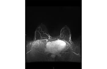
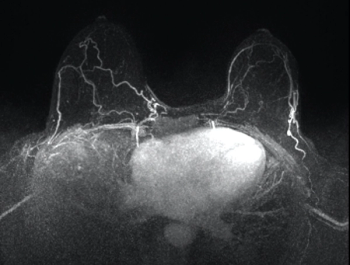
The value of GBCA in MRI images
Author(s)Ilana Cass, MD, Rola Saouaf, MD
These ten images can help ob/gyns recognize the additional information GBCA administration provides in MRIs.
Advertisement
Advertisement
Related Content


Advertisement
Advertisement
Trending on Contemporary OB/GYN
1
FDA: Levonorgestrel/ethinyl estradiol birth control patch sees update to labeled strength
2
Spotlighting menopause and mental health with Jessica Gaulton, MD, MPH
3
Using a self-collected vaginal fluid test for endometrial cancer detection
4
Shared decision-making key to non-hormonal VMS and sleep management in menopause
5