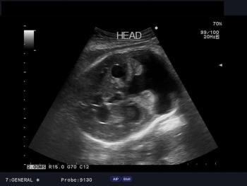
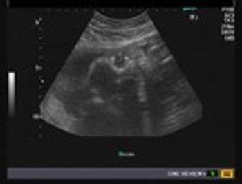
Identify the anomaly in this 35 week fetus as seen in these axial and sagittal images of fetal head.
Identify the anomaly in this 35 week fetus as seen in these axial and sagittal images of fetal head.
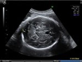
This patient had a history of pelvic pain and five weeks amenorrhea with no h/o spotting. What is your diagnosis?
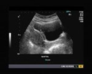
What is your diagnosis in this 40 year-old female patient with severe suprapubic pain exacerbated during menses? Ovaries were normal.
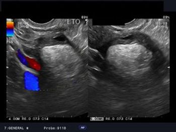
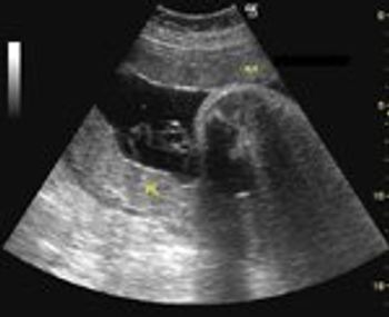
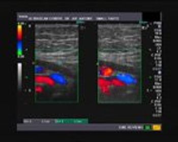
What is your diagnosis based on these images of the left adnexa? The patient is a young woman with pain in the same region.
This is the case of a pregnancy at 12 weeks gestational age. The patient underwent routine prenatal transabdominal and transvaginal ultrasound imaging.
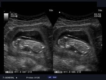
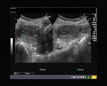
Test your diagnostic skills. What is your diagnosis of these endocavity scans?
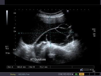
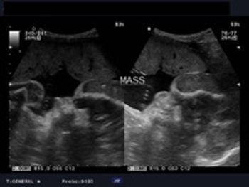
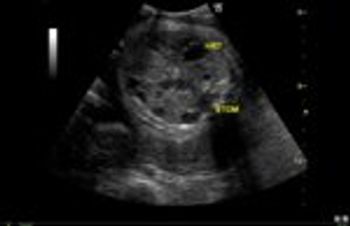
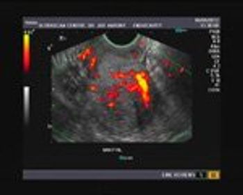
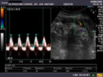
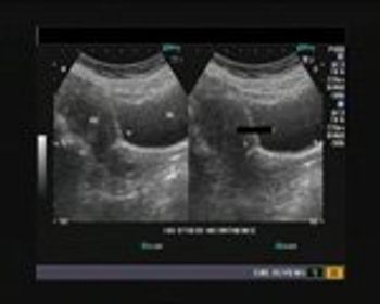
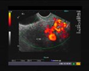
These ultrasound images show a right ovarian lesion.
These are ultrasound images of an early pregnancy.
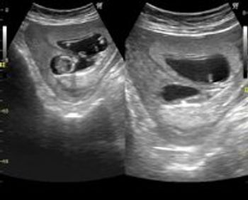
Our patient presented at 29 weeks gestation for a routine prenatal ultrasound.
There are multiple anomalies in these ultrasound images of a second trimester pregnancy.
This is an ultrasound scan of the placenta.
Our patient is a 30 year old female with a history of mild pain during micturition.
This is an ultrasound scan of the pelvis in 35 year old female patient.
Using only this image, describe what phase of the menstrual cycle this patient is in.
This is a normal 3 vessel view of a 34 week old fetal heart.
Our patient presented at 30 weeks gestation for a routine prenatal ultrasound.
What is your diagnosis of this 3rd trimester ultrasound?
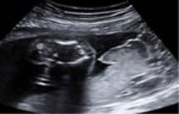
Our patient presents with 6 weeks of amenorrhea. What's your diagnosis?
In this case we will observe a 27 year old female patient with a history of polymenorrhea. What is your diagnosis of her ultrasound images?
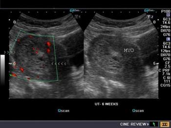
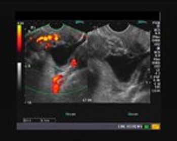
Our patient underwent a medical abortion at 5 weeks. What are the possible diagnoses for these images?
This is our LSCS patient, post-op. Currently, she has a history of pain in the lower abdomen and pelvis with scanty discharge from wound site.
This is a 30 year old female with polymenorrhea with dysmenorrhoea. What do the ultrasound images show?
This is a case of a female patient presenting with a history of 5 weeks of amenorrhoea and scanty bleeding. She was sent for diagnostic ultrasound imaging.
These are ultrasound images from a normal 3rd trimester fetus. What fetal anatomy do you see?
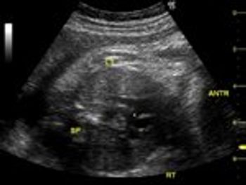
These ultrasound images show right hydroureter with hydronephrosis. What else do you see and what is the cause?
This is the case of a routine ultrasound examination at 31 weeks gestation.
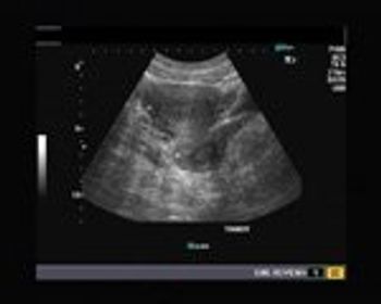
What is your diagnosis of this ultrasound image?
Test your ob/gyn ultrasound knowledge in our DailyDx.
Test your diagnostic skills!
This was a female patient 32 years old, who had irregular menses. She was married for 5 years but unable to conceive.